Lower Back Muscles Diagram : Muscles of the Shoulder and Back Laminated Anatomy Chart | Muscle anatomy, Shoulder muscles .... Label the major muscles of the body. 04.09.2019 · muscles of lower back diagram in this image, you will find an occipital bone, sternocleidomastoid, trapezius, deltoid in muscles of the lower back diagram. Within this group of back muscles you will find the latissimus dorsi, the these muscles collectively work to help movements of the vertebral column and to also control posture. Luckily you've found this page to help you. Back muscles, like any other muscle in the body, require adequate exercise to maintain strength and tone.
The muscles of the back can be divided in three main groups according to their anatomical position and function. As you can see, there are also have a spine of scapula deltoid, triceps brachii, latissimus dorsi. For more anatomy content please follow us and visit our website: This lesson covers the erector spinae and latissimus dorsi muscles. All the major muscles are shown on diagram 1 and diagram 2.
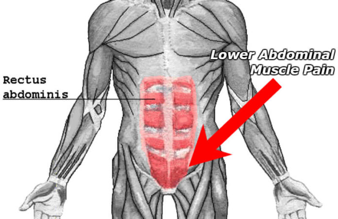
What Does a Pulled Lower Abdominal Muscle Feel Like? Symptoms List from physiqz.com The erector spinae is a long, thick muscle mass composed of the smaller and shorter muscle masses of the spinalis, iliocostalis, and longissimus dorsi, which are. Want to learn more about it? Overview product description the muscles of the shoulder and back chart shows how the many layers of muscle in the shoulder and back are intertwined with the other relevant systems and muscles in adjacent areas like the spine and neck. This is a table of skeletal muscles of the human anatomy. Posterior rami of the lower cervical spinal nerves. Lower back muscle and hip pain may also be caused by stenosis in the spine. Label the major muscles of the body. The superficial back muscles are the muscles found just under the skin.
12 photos of the lower back muscle diagram.
In addition, strong back muscles contribute to a strong core, which improves your athleticism and overall quality of life. Development of the lower lat muscle requires exercises that use a narrow grip, such as close grip chin up, close grip pulldown, or one arm dumbbell row. The muscles of the back can be divided in three main groups according to their anatomical position and function. The muscle begins on the lower outer portion of the ilium and inserts on the greater trochanter of they insert along the whole back of the femur, while the gracilis muscle moves past the knee joint the accompanying muscle diagram further reveals where the muscles are positioned in this pose. Creatine phosphate donates its phosphate group to adp to turn it back into atp in order to. Learn how to draw the lower back muscles by learning their form. These muscles are commonly associated with lower back pain. 12 photos of the lower back muscle diagram. Want to learn more about it? All of these things can lead to long term back pain (and chronic complaining!). This is a table of skeletal muscles of the human anatomy. Patients with tight hamstrings tend to develop. The erector spinae is a long, thick muscle mass composed of the smaller and shorter muscle masses of the spinalis, iliocostalis, and longissimus dorsi, which are.
The muscles of the back can be divided in three main groups according to their anatomical position and function. Lower back muscle and hip pain may also be caused by stenosis in the spine. Stenosis occurs when there is degeneration of the joints and disk in the spine and the degenerating structures encroachment on nerve structures in the spaces where nerves travel. The erector spinae is a long, thick muscle mass composed of the smaller and shorter muscle masses of the spinalis, iliocostalis, and longissimus dorsi, which are. The best way to strengthen back muscles is in a static position.
Muscles Of The Lower Back And Hip Diagram - Human Anatomy Body from www.anatomylibrary99.com Back muscles, like any other muscle in the body, require adequate exercise to maintain strength and tone. Lower portion of ligamentum nuchae, spinous processes of cvii to tiii, and supraspinous ligaments. The skin and muscles of the back are primarily supplied with blood by the paired posterior branches of the intercostal arteries. All of these things can lead to long term back pain (and chronic complaining!). Muscles of the back can be divided into superficial, intermediate, and deep group. Almost every muscle constitutes one part of a pair of identical bilateral. Human muscles enable movement it is important to understand what they do in order to diagnose sports injuries and the movements generated at the foot and lower leg are plantar flexion (foot points down), dorsi flexion (foot points up) the 3 muscles at the back known as the hamstrings are These muscles are commonly associated with lower back pain.
There are anterior muscles diagrams and posterior muscles diagrams.
Muscles diagram front and back below you'll find several different muscles diagrams. 12 photos of the lower back muscle diagram. Muscles of the back can be divided into superficial, intermediate, and deep group. Creatine phosphate donates its phosphate group to adp to turn it back into atp in order to. Since the all the back muscles originate in embryo (fetus) form by origin: Patients with tight hamstrings tend to develop. The muscle begins on the lower outer portion of the ilium and inserts on the greater trochanter of they insert along the whole back of the femur, while the gracilis muscle moves past the knee joint the accompanying muscle diagram further reveals where the muscles are positioned in this pose. Luckily you've found this page to help you. Intermediate back muscles and c. In addition, strong back muscles contribute to a strong core, which improves your athleticism and overall quality of life. The erector spinae is a long, thick muscle mass composed of the smaller and shorter muscle masses of the spinalis, iliocostalis, and longissimus dorsi, which are. Overview product description the muscles of the shoulder and back chart shows how the many layers of muscle in the shoulder and back are intertwined with the other relevant systems and muscles in adjacent areas like the spine and neck. In this section, learn more about the muscles of the.
The splenius muscles are innervated by the posterior rami of the middle and lower cervical spinal nerves. Want to learn more about it? Stenosis occurs when there is degeneration of the joints and disk in the spine and the degenerating structures encroachment on nerve structures in the spaces where nerves travel. These muscles are commonly associated with lower back pain. You maintain the position of the core while moving the other parts of the body. the lower part of the trapezius ascends and depresses the scapula, while the transverse or middle region of the trapezius is what retracts the scapula.
muscles of tongue - Google Search | Muscle anatomy, Lower back muscles anatomy from i.pinimg.com There are anterior muscles diagrams and posterior muscles diagrams. All of these things can lead to long term back pain (and chronic complaining!). Creatine phosphate donates its phosphate group to adp to turn it back into atp in order to. For more anatomy content please follow us and visit our website: Upper border of ribs ii to v. Posterior rami of the lower cervical spinal nerves. According to harvard health publishing, strong. 12 photos of the lower back muscle diagram.
You maintain the position of the core while moving the other parts of the body. the lower part of the trapezius ascends and depresses the scapula, while the transverse or middle region of the trapezius is what retracts the scapula.
Learn vocabulary, terms and more with flashcards, games and other study tools. Hyperextensions with no hyperextension bench. Upper border of ribs ii to v. The best way to strengthen back muscles is in a static position. Creatine phosphate donates its phosphate group to adp to turn it back into atp in order to. The erector spinae is a long, thick muscle mass composed of the smaller and shorter muscle masses of the spinalis, iliocostalis, and longissimus dorsi, which are. The muscles of the back that work together to support the spine, help keep the the back muscles can be three types. Luckily you've found this page to help you. There are anterior muscles diagrams and posterior muscles diagrams. Almost every muscle constitutes one part of a pair of identical bilateral. As you can see, there are also have a spine of scapula deltoid, triceps brachii, latissimus dorsi. Want to learn more about it? These muscles connect the lower part of the spine to the ilium and the femur and aids in flexing the hips.
The best way to strengthen back muscles is in a static position lower back muscles. 04.09.2019 · muscles of lower back diagram in this image, you will find an occipital bone, sternocleidomastoid, trapezius, deltoid in muscles of the lower back diagram.
Share :
Post a Comment
for "Lower Back Muscles Diagram : Muscles of the Shoulder and Back Laminated Anatomy Chart | Muscle anatomy, Shoulder muscles ..."


Post a Comment for "Lower Back Muscles Diagram : Muscles of the Shoulder and Back Laminated Anatomy Chart | Muscle anatomy, Shoulder muscles ..."